Avaliação e Monitoramento embrionário 24h
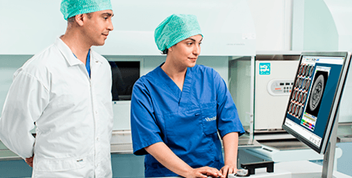
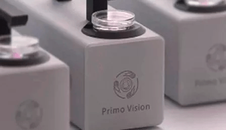
O Time-Lapse do embrião é um método de avaliação não-invasiva, possibilitando ao embriologista uma seleção criteriosa dos melhores embriões a serem transferidos para o útero. O PrimoVision™ é um equipamento que conta com uma câmera que monitora os embriões de dentro da incubadora 24 horas por dia. São geradas fotos, automaticamente, durante todo desenvolvimento dos embriões e um vídeo de todo o processo ao final do tratamento, sendo hoje o que há de mais moderno em Reprodução Humana Assistida no mundo.
Somos a única clínica da região e uma das dez do país a contar com essa tecnologia, beneficiando o tratamento de fertilização nos seguintes aspectos:
- Potencialização dos Resultados: as imagens de alta qualidade permitem a monitorização detalhada dos embriões e avaliações precisas, detectando alterações no desenvolvimento que não são identificadas pela microscopia convencional, como a divisão assíncrona e alterações morfológicas relevantes.
- Minimização do stress na cultura embrionária: o sistema de monitorização colocado no interior da incubadora evita a mobilização desnecessária dos embriões ou sua exposição à luz, gerando embriões mais saudáveis.
Seleção embrionária
Hoje, utilizamos a seleção do embrião para transferência baseada na morfologia no momento da avaliação. Com o monitoramento embrionário 24h PrimoVision™, a seleção se baseia também no desenvolvimento dos embriões. Ou seja, não vemos somente o momento atual da seleção, mas temos acesso à toda cinética embrionária, vendo como o embrião passou por todos os processos desde a fertilização até o momento em que estamos escolhendo aquele com maior potencial de gestação.
O software utilizado, baseado nas marcações estipuladas pelos especialistas, mostra quais embriões são mais aptos à transferência. Desse modo, podemos somar à análise morfológica a análise do desenvolvimento do embrião para escolher aquele com maior potencial de implantação. Além disso, os médicos e embriologistas tem acesso às imagens dos embriões que estão sendo monitorados pelo PrimoVision™, pois o equipamento permite a visualização através do celular ou tablet.

Marcelo Rufato
CRBio 64065/01
Embriologista e diretor dos Laboratórios do CEFERP.
- Basile N, Vime P, Florensa M, Aparicio Ruiz B, García Velasco JA, Remohí J, Meseguer M. (2015) “The use of morphokinetics as a predictor of implantation: a multicentric study to define and validate an algorithm for embryo selection” Hum Reprod. 2015 Feb;30(2):276-83
- Bodri D, Kato R, Kondo M, Hosomi N, Katsumata Y, Kawachiya S, Matsumoto T. (2015) “Time-lapse monitoring of zona pellucida-free embryos obtained through in vitro fertilization: a retrospective case series” Fertil Steril. 2015 Mar 12. pii: S0015-0282(15)00138-7
- Bronet F, Nogales MC, Martínez E, Ariza M, Rubio C, García-Velasco JA, Meseguer M. (2015) ”Is there a relationship between time-lapse parameters and embryo sex?” Fertil Steril. 2015 Feb;103(2):396-401.e2
- Cetinkaya M, Pirkevi C, Yelke H, Colakoglu YK, Atayurt Z, Kahraman S. (2015) “Relative kinetic expressions defining cleavage synchronicity are better predictors of blastocyst formation and quality than absolute time points” J Assist Reprod Genet. 2015 Jan;32(1):27-35
- Chamayou S, Romano S, Alecci C, Storaci G, Ragolia C, Palagiano A, Guglielmino A. (2015) “Oocyte vitrification modifies nucleolar remodeling and zygote kinetics-a sibling study” J Assist Reprod Genet. 2015 Apr;32(4):581-6
- Chawla M, Fakih M, Shunnar A, Bayram A, Hellani A, Perumal V, Divakaran J, Budak E. (2015) “Morphokinetic analysis of cleavage stage embryos and its relationship to aneuploidy in a retrospective time-lapse imaging study” J Assist Reprod Genet. 2015 Jan;32(1):69-75
- Fréour T, Le Fleuter N, Lammers J, Splingart C, Reignier A, Barrière P. (2015) “External validation of a time-lapse prediction model” Fertil Steril. 2015 Apr;103(4):917-22
- Milewski R, Kuć P, Kuczyńska A, Stankiewicz B, Łukaszuk K, Kuczyński W. (2015) “A predictive model for blastocyst formation based on morphokinetic parameters in time-lapse monitoring of embryo development” J Assist Reprod Genet. 2015 Apr;32(4):571-9
- Mölder A, Drury S, Costen N, Hartshorne GM, Czanner S. (2015) “Semiautomated analysis of embryoscope images: Using localized variance of image intensity to detect embryo developmental stages” Cytometry A. 2015 Feb;87(2):119-28
- Park H, Bergh C, Selleskog U, Thurin-Kjellberg A, Lundin K. (2015) “No benefit of culturing embryos in a closed system compared with a conventional incubator in terms of number of good quality embryos: results from an RCT” Hum Reprod. 2015 Feb;30(2):268-75
- Stensen MH, Tanbo TG, Storeng R, Åbyholm T, Fedorcsak P. (2015) ”Fragmentation of human cleavage-stage embryos is related to the progression through meiotic and mitotic cell cycles” Fertil Steril. 2015 Feb;103(2):374-81.e4
- Aguilar J, Motato Y, Escribá MJ, Ojeda M, Muñoz E, Meseguer M. (2014) “The human first cell cycle: impact on implantation” Reprod Biomed Online 28(4):475-484
- Basile N., Nogales, M.C., Bronet, F., Florensa, M., Riqueiros, M., Rodrigo, L., Garcia-Velasco, J., Meseguer, M. (2014) ”Increasing the probability of selecting chromosomally normal embryos by time-lapse morphokinetic analysis” Fertil Steril 2014; 101(3), 699-704
- Campbell A, Fishel S, Laegdsmand M. (2014) “Aneuploidy is a key causal factor of delays in blastulation: author response to ‘A cautionary note against aneuploidy risk assessment using time-lapse imaging” Reprod Biomed Online 28(3): 279-83
- Desai N, Ploskonka S, Goodman LR, Austin C, Goldberg J, Falcone T (2014) “Analysis of embryo morphokinetics, multinucleation and cleavage anomalies using continuous time-lapse monitoring in blastocyst transfer cycles” Reprod Biol Endocrinol 12:54 doi: 10.1186/1477-7827-12-54
- Ergin EG, Çalişkan E, Yalçinkaya E, Öztel Z, Çökelez K, Özay A, Özörnek HM (2014) ”Frequency of embryo multinucleation detected by time-lapse system and its impact on pregnancy outcome” Fertil Steril 102(4): 1029–1033
- Joergensen MW, Agerholm I, Hindkjaer J, Bolund L, Sunde L, Ingerslev HJ, Kirkegaard K. (2014) ”Altered cleavage patterns in human tripronuclear embryos and their association to fertilization method: A time-lapse study” J Assist Reprod Genet 31(4): 435-42
- Lammers J, Splingart C, Barriére P, Freour T. (2014) “Morphokinetic parameters of ICSI tripronucleated embryos observed using time lapse” Reprod Biomed Online 28(5): 658-60
- Li R, Pedersen KS, Liu Y, Pedersen HS, Lægdsmand M, Rickelt LF, Kühl M, Callesen H (2014) ”Effect of red light on the development and quality of mammalian embryos” J Assist Reprod Genet 31(7): 795-801
- Liu Y, Chapple V, Roberts P, Ali J, Matson P. (2014) “Time-lapse videography of human oocytes following intracytoplasmic sperm injection: events up to the first cleavage division” Reprod Biol. 2014 Dec;14(4):249-56
- Liu Y, Chapple V, Roberts P, Matson P (2014) “Prevalence, consequence, and significance of reverse cleavage by human embryos viewed with the use of the Embryoscope time-lapse video system” Fertil Steril doi: 10.1016/j.fertnstert.2014.07.1235
- Kirkegaard K, Campbell A, Agerholm I, Bentin-Ley U, Gabrielsen A, Kirk J, Sayed S, Ingerslev HJ (2014) “Limitations of a time-lapse blastocyst prediction model: a large multicentre outcome analysis” Reprod Biomed Online 29(2):156-158
- Morbeck, D, Paczkowski M, Fredrickson JR, Krisher RL, Hoff HS, Baumann NA, Moyer T, Matern D (2014) “Composition of protein supplements used for human embryo culture” J Assist Reprod Genet, doi: 10.1007/s10815-014-0349-2
- Morbeck DE, Krisher RL, Herrick JR, Baumann NA, Matern D, Moyer T (2014) ” Composition of commercial media used for human embryo culture” Fertil Steril 102(3): 759-766
- Rienzi L, Capalbo A, Stoppa M, Romano S, Maggiulli R, Albricci L, Scarica C, Farcomeni A, Vajta G, Ubaldi FM (2014) “No evidence of association between blastocyst aneuploidy and morphokinetic assessment in a selected population of poor-prognosis patients: A longitudinal cohort study” Reprod Biomed Online DOI: http://dx.doi.org/10.1016/j.rbmo.2014.09.012
- Rubio I,Galán A, Larreategui Z, Ayerdi F, Bellver J, Herrero J, Meseguer M (2014) “Clinical validation of embryo culture and selection by morphokinetic analysis: a randomized, controlled trial of the EmbryoScope” Fertil Steril. DOI: http://dx.doi.org/10.1016/j.fertnstert.2014.07.738
- Stecher A, Vanderzwalmen P, Zintz M, Wirleitner B, Schuff M, Spitzer D, Zech NH (2014) “Transfer of blastocysts with deviant morphological and morphokinetic parameters at early stages of in-vitro development: a case series” Reprod Biomed Online 28(4): 424-435
- Wilken-Jensen HN, Kristensen SG, Jeppesen JV, Yding Andersen C (2014) ”Developmental competence of oocytes isolated from surplus medulla tissue in connection with cryopreservation of ovarian tissue for fertility preservation” Acta Obstet Gynecol Scand 93(1):32-7
- Wissing ML, Bjerge MR, Olesen AI, Hoest T, Mikkelsen AL. (2014) “Impact of PCOS on early embryo cleavage kinetics” Reprod Biomed Online 28(4): 508-14
- Yang Z, Zhang J, Salem SA, Liu X, Kuang Y, Salem RD, Liu J (2014) “Selection of competent blastocysts for transfer by combining time-lapse monitoring and array CGH testing for patients undergoing preimplantation genetic screening: a prospective study with sibling oocytes” BMC Medical Genomics 2014, 7:38